Neuroimaging – An Introduction to How We Measure Brain Performance
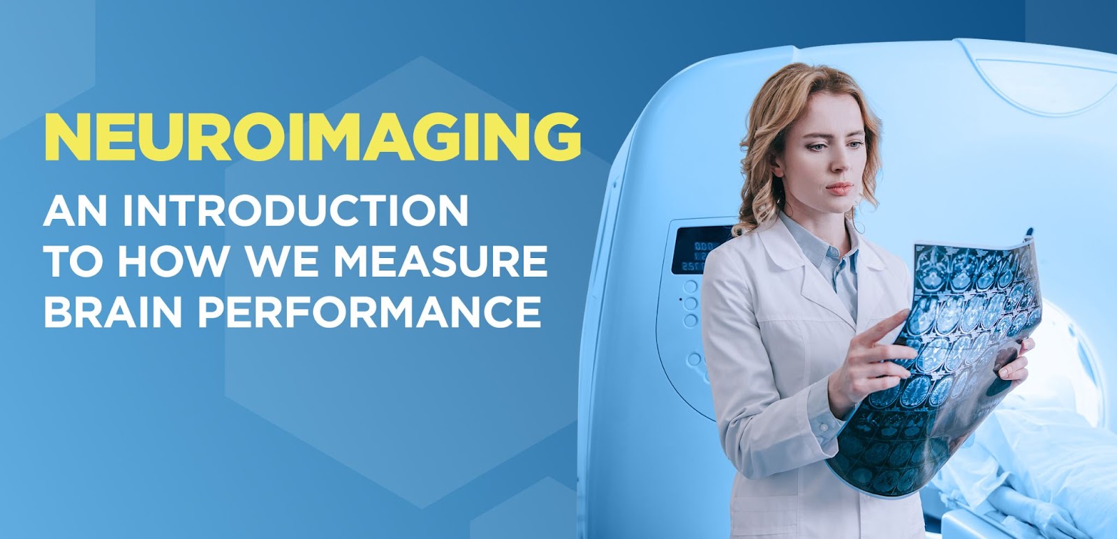
Table of Contents:
- What is Neuroimaging
- How Can Neuroimaging Help Diagnose a Problem
- What are the Different Neuroimaging Options Available Today?
- CT/CAT
- MRI
- EEG
- EROS
- Additional Tests to Determine Brain Health
- WAVi
- PET Scan
- SPECT Scan
- Blood, Urine, or Other Bodily Fluids
- Genetic Testing
- IQ Tests
- How Clarke Bioscience Uses Neuroimaging to Help Understand our Customers
- Conclusion
The brain is a mysterious and complicated organ. It’s full of sensitive structures and complex elements that help manage the entire body. Taking a peek under the hood can offer valuable insight into its current health but getting a visual of the brain is not an easy task. This organ is fortified by a shield of bone, fluid, and a roadmap of blood vessels that keep it safe.
This is where one of medicine’s best advancements comes in: brain-imaging tests.
Brain imaging tests (or neuroimaging) provide medical professionals with a direct view of the brain without cutting into the body. Many of these imaging tests even allow the functions of the brain to be seen in real-time. These inventions have remained unmatched for helping doctors and scientists understand the way the brain works.
This article discusses neuroimaging and how it can be used to diagnose diseases and help save lives. Then, we talk about the different types of brain imaging tests currently available and the incredible information they provide.
What Is Neuroimaging?
Let’s increase the font size here to match with the other images
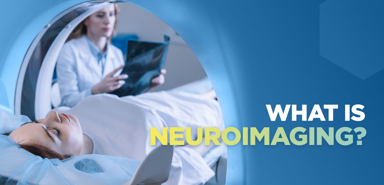
As the name suggests, neuroimaging is a form of medical imaging that focuses on the brain’s neurological form and function. Neuroimaging is routinely conducted in both healthy and sick populations to understand the way the brain works. Because of its availability, neuroimaging has helped neuroscientists, psychologists, psychiatrists, basic scientists, and other clinicians advance their understanding of their patient’s brains at a very rapid pace.
How Can Neuroimaging Help Diagnose a Problem?
This needs to read “How Can It Help?”
Before neuroimaging was available, medicine still had a general idea of the way the brain worked. Doctors and scientists could use “barbaric” procedures, exploratory surgery, and autopsy to understand neurological diseases and disorders. But this basic approach wasn’t enough to create effective treatments. In fact, just three decades ago, medical students were still hesitant to go into the field of neurology because of the lack of treatment options that existed (1). Neuroimaging changed all of that. With this advancement, researchers were granted a direct view into the brain and could see active tumors, bleeds, and damage without interference from other structures.
What are the Different Neuro Imaging Options Available Today?
The introduction of noninvasive neuroimaging in the early 1900s started a landslide of inventions. Today there are many different options for examining the brain, and a few of them even allow it to be viewed in real-time. Here are a few of them:
CT/CAT Scan
The CT (Computed Tomography) scan was the very first invention that allowed for detailed pictures of the brain. It was developed in 1972 by Godfrey Hounsfield, an engineer with no history in the medical field, and was revolutionary (2). Hounsfield went on to win the Nobel Peace Prize in 1979 for his discovery.
A CT scan uses technology like an X-Ray to produce lots of different 2-D images of the brain. Unlike an X-ray, CT scans contain a beam that moves around the body in a circular motion. This allows doctors to view the brain from lots of different angles. Sometimes, “contrast” or dye can be administered through an IV to help enhance the brain images, allowing different brain structures to stand out during the scan.
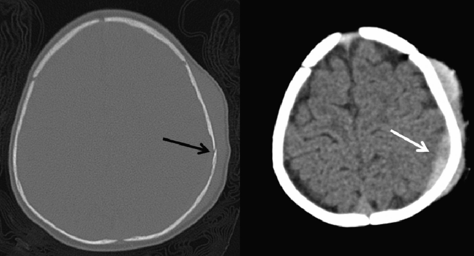
Image Credit: American Journal of NeuroRadiology3
Above, you will see two photos from a CT scan taken on a 5-month-old child after falling 3-4 feet and landing on a wooden floor. The black arrow shows a minor fracture of the skull and the white one points to a collection of fluid that resulted from the fall.
Brain CT Scans continue to be used in most hospitals today and typically take about 30-60 minutes to complete. They provide clinicians with images of the bone, soft tissue, and blood vessels simultaneously, allowing them to gain a detailed view of the brain. CT Scans of the head are used to identify and diagnose tumors, bleeding, structural abnormalities, and even infection. They are often used to investigate head and neck injuries and assess damage when a patient comes in with a stroke. CT Scans are most beneficial when other types of assessments, like X-rays or physical examination, are inconclusive.
MRI
CT Scans were not the only imaging advancement in 1972. The MRI was also invented that same year by American physician Raymond Damadian (9). Damadian developed the scanner because he believed it would be helpful to detect cancer in humans.
Unlike CT Scans though, images produced by MRI are three-dimensional. The machine uses not radiation but magnets to detect and monitor the brain and its function. MRI scanners look very similar to CT machines, and the patient must remain very still during the procedure.
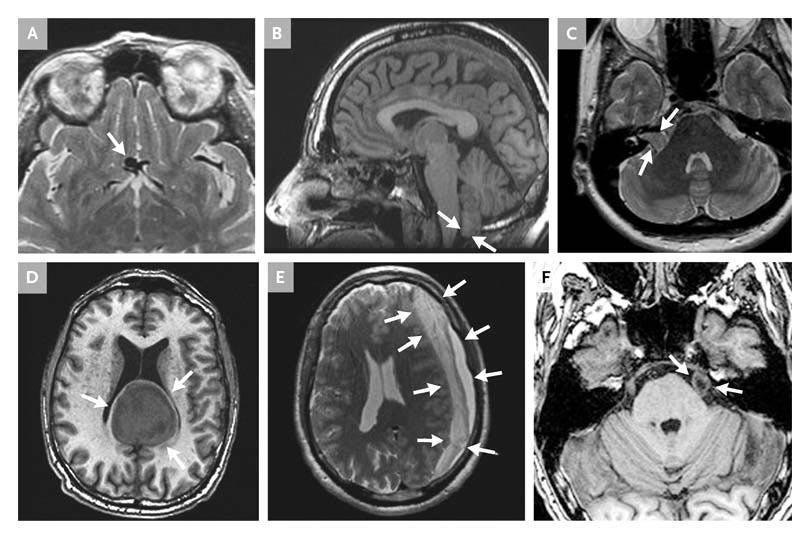
Image Credit: The New England Journal of Medicine (10)
Above, you will see several different incidental findings on MRI scans of the brain. The arrows indicate the abnormalities, which are as follows:
- Panel A: Aneurysm
- Panel B: Chiari malformation
- Panel C: Noncancerous tumor
- Panel D: Noncancerous tumor
- Panel E: Brain bleed
- Panel F: Noncancerous tumor
An MRI of the brain typically takes 30-60 minutes to complete and can be helpful any time a doctor needs to get a look inside someone’s head. This type of scan can be used to detect brain tumors, brain injuries, and even the causes of headaches.
And remember, MRI is not the same as CT.
Besides their apparent differences, MRI has additional capabilities. On top of taking static images of the brain, fMRI (functional MRI) can monitor its function. This type of test can assess damage to the brain and how it might affect a person’s life. A patient is usually asked to complete a task (evoked response potential) with this type of test while the brain’s function is monitored.
DTI (Diffusion Tensor Imaging) is another technique used in MRI and is mainly used to map the brain before surgery. It estimates the location of different white matter tracts by detecting levels of water in the brain. DTI imaging is significant because it can help surgeons preserve essential structures of the brain during surgery. DTI provides a 3D, high-resolution snapshot of a brain and its structure.
EEG
EEG is sometimes considered the father of neuroimaging techniques because it was the first, real noninvasive glimpse into the brain. The technology takes advantage of the electrical properties within the brain and notes the fluctuations within them. Human brain activity was first recorded through EEG by German psychiatrist Hans Berger in 1924 (11).
EEG is still used today and is especially helpful for detecting seizures. Through the electrodes that are secured to the scalp, abnormal brain patterns can be identified and explored. However, instead of producing an “image,” as you might expect from an MRI or CT Scan, EEG sends signals to a computer to form patterns.
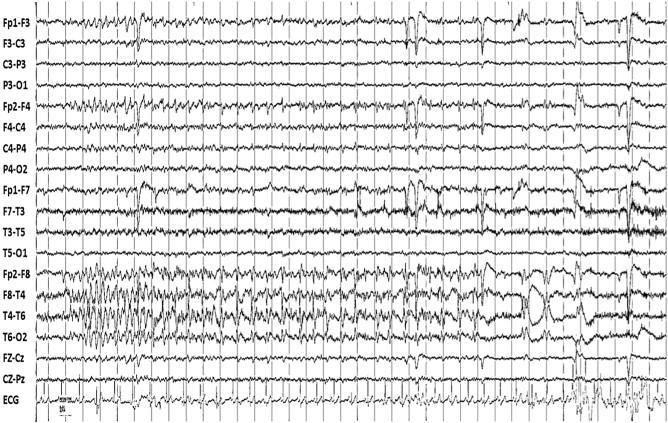
Image Credit: ScienceDirect12
Above, you will see a seizure recorded in a child through an EEG. Most EEG procedures take approximately 1 hour but can sometimes last longer.
EROS
Event-related Optical Signal (EROS) is one of the newest technologies available in brain imaging. Developed at the University of Illinois in 1995, this noninvasive tool also uses light and optical fibers to measure the function of the brain near the cerebral cortex.
In EROS scans, the light is infrared and can detect changes within the brain in milliseconds and focus on activity within millimeters (7).
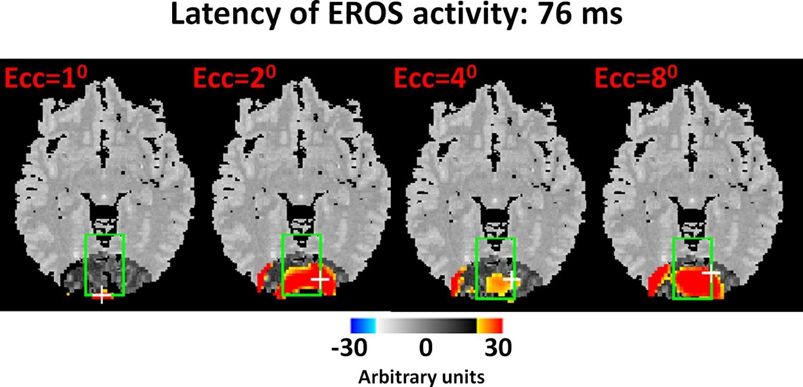
Image Credit: Frontiers in Neuroscience (8)
Above, you will see a 3D recreation of an EROS scan with different points of stimulation. Although these scans contain a high-quality image, they’re limited to detecting activity no more than a few centimeters deep. EROS may be beneficial for identifying issues with oxygenation in the brain, especially when related to migraine disorders and other neurological or psychological conditions. The EROS scan and its applications are still being researched.
Additional Tests to Determine Brain Health
Again, here I would like to increase the font size
Advancements for brain imaging are improving every year. Some additional brain imaging tests that yield results in a clinical setting include:
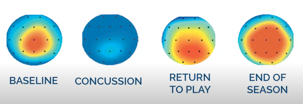
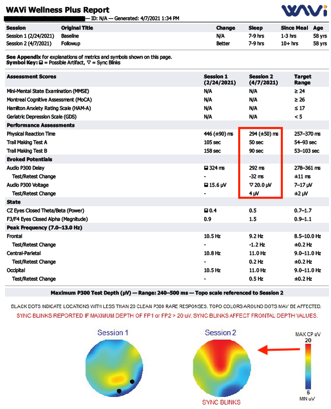
WAVi
A new, portable brain scan, WAVi, assesses the brain on multiple different levels. It uses ERP (Evoked Response Potential) and EEG testing to determine brain function and response to prescribed interventions.
The WAVi test provides a snapshot of your brain’s voltage (power), reaction time (speed), and connectivity. It also provides a color coded topogram of where your brain’s most active regions are and where you might be deficient. Lastly, it measures the four most measured brain waves (Beta, Alpha, Theta & Delta), which provide a glimpse into your state of mind.
Much research is being conducted on the importance of these biomarkers and how they can help diagnose an injury or disease and track the recovery process when using a prescribed intervention.
PET scan
A PET (Positron Emission Tomography) scan uses a specialized radioactive dye (tracer) to check for diseases throughout the body. The dye is absorbed by certain organs and tissues and allows doctors to see how well they’re working. In brain disorders, PET scans can measure blood flow, oxygen use, central nervous system abnormalities, and most importantly, how the brain is processing glucose.
PET Scans are great because they can show changes at the cellular level, unlike CT Scans and MRIs. Since everything begins at the cellular level, you can identify potential issues before changes occurring in the organs or tissues are noticeable by a CT or MRI scan.
SPECT Scan
Like an MRI, SPECT (Single-Photon Emission Computerized Tomography) scans use nuclear imaging to provide a 3D depiction of your brain. This test integrates the CT scan and a radioactive tracer. This tracer emits Gamma rays and displays them on the CT cross-sections.
It allows the physician to see the function of your organs. SPECT Scans are often used for assessing patients with dementia, seizures, and epilepsy.
Blood, Urine, or Other Bodily Fluids
Blood and urine testing is an essential step in any brain testing. Testing bodily fluids can help scientists understand the severity of a disease, and it can also be beneficial for monitoring levels of medication in the body. Specific genetic identifiers can also be picked up in the blood and salivary cells. These include indicators for epilepsy, encephalitis, and meningitis.
Genetic Testing
As mentioned above, lots of different genetic components have been identified in certain brain disorders. And science is getting better at recognizing these. Genetic testing is becoming a regular step in every healthy pregnancy. By taking the mother’s blood, scientists can test an unborn child for issues like Down Syndrome and even epilepsy.
IQ Tests
While IQ testing is often viewed as a fun way to see how “smart” you are, this basic assessment can also be used to determine brain function. IQ tests can be beneficial for identifying intellectual disabilities and cognitive functioning. However, the IQ test should never be the only way of determining healthy brain function.
How Dr. Clarke and Clarke Bioscience Use Brain Imaging to Help Understand Our Customers
At Clarke Bioscience, we recognize the tangible value that cutting-edge technology has on the health of the world. We combine nutrition, biochemical formulations, and advanced diagnostic imaging to provide the most support for your brain. Imaging solutions help us understand you, which only increase the efficacy of our products.
We currently use a mixture of imaging tests but utilize the WAVi most consistently to understand the power, speed and connectivity of a subject’s brain. This has helped us immensely by providing objective feedback to our customers when they take one of Dr. Clarke’s interventions.
This test is fast, noninvasive and provides Dr. Clarke with a clear picture of where you stand before and after an intervention.
Conclusion
Viewing the brain in action and at rest has provided humankind with an understanding like never before. We finally realize the deep connections that the brain has with every part of the body, including our own emotions and thoughts.
This is why brain imaging is so important. It helps us understand concepts like never before so that we can take charge of our health. Leading a life of optimal health starts with you, and we’re ready to take it with you.
References
- Valeo, T. (2013). New Brain-Imaging Techniques Help Diagnose Neurologic Conditions. Brain&Life. https://www.brainandlife.org/articles/new-brain-imaging-techniques-provide-better-ways-to-diagnose-and/
- Ambrose, R. (n.d.). Godfrey Hounsfield. Radiopaedia. https://radiopaedia.org/articles/godfrey-hounsfield?lang=us
- Linscott, L. (2013, June). CT for Pediatric, Acute, Minor Head Trauma: Clinician Conformity to Published Guidelines. AJNR. http://www.ajnr.org/content/34/6/1252
- Boas, D. (2007, April 27). Near infrared imaging. Scholarpedia. http://www.scholarpedia.org/article/Near_infrared_imaging
- Wai Lee, C. (2017, May 31). Diffuse optical tomography to investigate the newborn brain. Pediatric Research. https://www.nature.com/articles/pr2017107/figures/4
- Hernandez-Martin, E. (2021, Feb 28). Diffuse optical tomography in the human brain: A briefly review from the neurophysiology to its applications. SAGE Journals. https://journals.sagepub.com/doi/full/10.26599/BSA.2020.9050014#
- Gratton, G. (1998, December). Dynamic brain imaging: Event-related optical signal (EROS) measures of the time course and localization of cognitive-related activity. SpringerLink. https://link.springer.com/article/10.3758/BF03208834
- Gratton, G. (2010, June 23). Fast optical imaging of human brain function. Frontiers in Neuroscience. https://www.frontiersin.org/articles/10.3389/fnhum.2010.00052/full
- Sharma, R. (n.d.). Raymond V Damadian. Radiopaedia. https://radiopaedia.org/articles/raymond-v-damadian?lang=us#
- Vernooij, M. (2007, Nov 1). Incidental Findings on Brain MRI in the General Population. The New England Journal of Medicine. https://www.nejm.org/doi/full/10.1056/nejmoa070972
- NCBI. (2016). https://www.ncbi.nlm.nih.gov/books/NBK390348/.
- Rizvi, S. (2013). Outpatient ambulatory EEG as an option for epilepsy surgery evaluation instead of inpatient EEG telemetry. ScienceDirect. https://www.sciencedirect.com/science/article/pii/S221332321300011X#f0005